


By Dr Oliver Liyou BVSc (Hons) MACVSc (Eq Dent)
www.evds.net.au
Introduction
The word Melanoma strikes fear into most Australians, given the harsh UV conditions we live in and the potential fatal outcome in humans who are affected with melanoma. However melanomas in horses are quite different, as they are usually benign and are not even induced by UV rays from the sun!
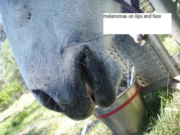
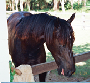
Melanomas on the lips & face.
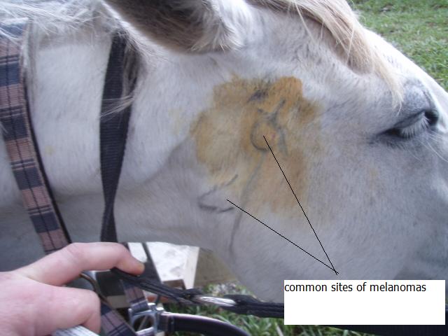
Sites of melanoma on side of face
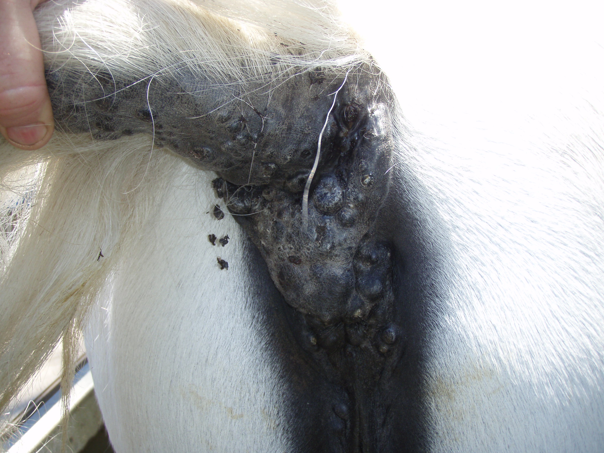
Clusters of melanomas under tail base.
What are they?
Melanomas in horses are brown or black nodules which start as small lumps in or just under the skin of horses, and slowly grow larger – sometimes. They can be hard, soft, solitary or appearing in clusters. Diagnosis of them is confirmed by doing a fine needle aspirate of the lump and examining it on a microscope slide. This simple technique is sometimes not enough for accurate diagnosis, as sometimes the melanoma tumour is non pigmented (amelanotic) and a proper tissue biopsy is required.
What horses are affected?
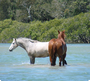
Most horses who contract melanomas are grey in colour, with the associated black skin (thus has more melanin pigment containing cells) under the grey hair. Approximately 80 % of grey horses over 9 years of age will get at least 1 melanoma tumour. The grey horse’s skin is quite different to the white hair coat and pink skin commonly seen on paint coloured horses, horses with large blazes and white socks etc. However many horses that start out with grey hair coat colour do become more white in hair colour with age, but their skin remains black through the ageing process.
Despite melanomas mainly being a problem in greys, non-grey horses can occasionally be affected. And in these horses, the tumour is usually more malignant and dangerous in nature.
Sites affected on horses
Melanomas are most commonly found under the tail base, but also can be found in the sheath skin and penis, around the eyelids, below the ears in the region of the parotid salivary gland, in the lips, as well as generally in the skin on the body. They can also spread to the lining of internal organs such as the liver, spleen, lungs and intestines, but this is a far less common presentation.
What causes them and how do they differ from Human Melanomas?
Human Melanomas are highly malignant tumours which are caused by excessive exposure to UV rays. However, in horses the cause is still unclear. It is known that they are a disturbance in the pigment production rate in the melanocytes (cells that produce pigment), but no one really knows why these cells become disturbed.
In horses they can develop in 3 basic ways:
1) They develop slowly over years, and remain that way for 10-20 years (luckily is the most common scenario)
2) They have existed for years in the benign state, and then suddenly turn malignant and spread rapidly to other parts of the body.
3) From the first time they appear, they are malignant and spread rapidly. Unfortunately this can even occur as a foal!
What can melanomas be confused with? The most common skin tumour of horses is the sarcoid tumour, which is a benign (slow growing and doesn’t usually spread to other organs) skin tumour and can take several different appearances. Other skin nodules include:
– Nodular necrobiosis – a non cancerous lump induced by biting insects.
– Squamous cell carcinoma – a malignant tumour induced by UV conditions, and thus usually affects areas of pink skin such as eyelids, muzzles, and penis regions.
Risks to horses with melanomas.
As the melanoma grows larger, they often cause problems through either ulcerating or becoming infected, or through interfering with normal functions in the body. For example a large melanoma growing beside the anus could make defecating increasingly difficult, or one beside the intestines could block off the bowel and cause a fatal colic.
The most challenging issue with melanomas is that , despite most of them being benign (and thus more of a cosmetic blemish than a health risk) , any one of them has the potential to one day turn malignant and invade other parts of the horse’s body and lead to a fatal outcome. Once a melanoma turns malignant, they often spread and appear as a spreading series of lumps, nodules or sheets of black tumour masses. At this point, they are very hard to cure, even with surgery.
Treatment Options.
As an owner of a grey horse, you must realise that from 9 years of age, you are 80% likely to find a melanoma on your horse. So then once you find one or more melanomas on your horse, you need to decide whether to either:
a) Play the odds and just monitor their size, and only have treatment done if they grow large or rapidly spread and expand.
b) Be very cautious and proactive and have them surgically excised when they are small (less than 3 cm) and easy to remove.
Treatments can involve one or a combination of:
1) Surgical excision – risks and success rates vary depending on the size and location.
2) Cryonecrosis – Freezing them off with liquid nitrogen – may need to repeat once or twice per year to “manage” them.
3) Chemotherapy – injecting drugs into the lesion on a continued or regular basis.
4) Radiation.
5) Cimetidine – a gastric ulcer treatment has been suggested but results vary and aren’t too encouraging.
6) Vaccine – some experimental evidence exists utilising an autogenous vaccine as treatment.